Dolor de rodilla: causas, síntomas y remedios
El dolor de rodilla es un trastorno común que afecta a personas de todas las edades. La «gonalgia», término médico que designa los síntomas dolorosos relacionados con la rodilla, puede deberse a una lesión, como la rotura del ligamento cruzado, o a la «reducción» del cartílago. Ciertas condiciones patológicas, incluida la artritis, también pueden causar dolor e inflamación de rodilla.
Hay muchos tipos de pacientes que acuden a una consulta de fisioterapia con un problema de rodilla: desde un paciente con una prótesis de rodilla hasta un deportista profesional con irritación crónica del tendón rotuliano.
Muchos tipos de dolor de rodilla responden bien a los tratamientos de fisioterapia y al uso de rodilleras que pueden ayudar a aliviar el dolor. En algunos casos, sin embargo, la rodilla puede requerir cirugía, por lo que siempre es una buena idea no subestimar el dolor.
La rodilla también tiene una estrecha relación funcional con las demás articulaciones de la extremidad inferior, por lo que siempre debe evaluarse en relación con ellas.
Índice:
- La rodilla: un poco de anatomía
- Síntomas y posibles causas de dolor de rodilla
- Remedios para el dolor de rodilla
- Información adicional: síndrome femororrotuliano
La rodilla: un poco de anatomía
La articulación de la rodilla es un ginglimo angular y se divide en 3 articulaciones:
– Articulación tibiofemoral
– Articulación patelofemoral (femororrotuliana)
– Articulación tibiofibular proximal
Debido a la forma de las superficies articulares, para una estabilidad funcional satisfactoria son necesarios los meniscos, el complejo ligamentoso capsular (ligamentos cruzados y colaterales) y un sistema muscular diferenciado.
Los ligamentos cruzados y colaterales guían y estabilizan la rodilla: garantizan que la posición de los cóndilos femorales en relación con la meseta tibial y los meniscos sea siempre la mejor.
Existen dos ligamentos colaterales:
- el ligamento colateral interno: ancho y conectado al menisco interno y a la cápsula.
- El ligamento colateral externo: con una forma más bien redonda, muy fuerte y sin conexión con el menisco interno.
Los principales músculos de la rodilla son:
– El cuádriceps femoral, formado por el recto femoral, el vasto intermedio, el vasto lateral y el vasto medial. Este último asume una función muy importante como estabilizador de la rótula, por lo que su rehabilitación desempeña un papel primordial en el síndrome patelofemoral.
– El compartimento posterior del muslo, formado por los músculos isquiocrurales (bíceps femoral, semimembranoso y semitendinoso).
– Los músculos aductores que forman la parte interna.
– El músculo tensor de la fascia lata en la parte externa del muslo.
Síntomas y posibles causas de dolor de rodilla
Los síntomas del paciente con dolor de rodilla pueden ser diferentes: el dolor puede producirse en la parte anterior (por debajo o por encima de la rótula), lateral/inferior o posterior (en la cavidad poplítea).
En algunos casos, el paciente puede experimentar un dolor que se extiende por «toda la rodilla».
El dolor de rodilla puede sentirse durante o después de un movimiento concreto: el paciente puede referir que el dolor se produce al caminar o correr, o al ponerse en cuclillas o mantener una posición flexionada durante mucho tiempo (por ejemplo, en el automóvil o en la mesa). A veces sólo se presenta al caminar cuesta abajo o al bajar escaleras. También puede haber hinchazón y pérdida de movilidad.
Las posibles causas de dolor de rodilla, en ausencia de una lesión por esguince o contusión, son:
– Síndrome femororrotuliano
– Síndrome de la cintilla iliotibial
– Bursitis (bursitis de la pata de ganso, prerrotulatoria o infrarrotulatoria)
– Desgaste del menisco
– Quistes de Baker
– Inflamación del cuerpo de Hoffa
– Rigidez articular (también como consecuencia de una intervención quirúrgica)
– Retracciones/Puntos gatillo en el cuádriceps
– Tendinopatía distal de los isquiotibiales
– Rigidez del tobillo
– Apoyo incorrecto del pie
Remedios para el dolor de rodilla
En el caso de una lesión de esguince de rodilla, que provoca dolor e hinchazón, el primer paso es consultar a un Traumatólogo para obtener un diagnóstico correcto. El médico realizará pruebas clínicas y posiblemente prescribirá pruebas diagnósticas (como la resonancia magnética) para llegar a un diagnóstico correcto.
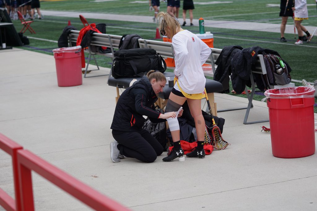
En estos casos, en la fase inicial, se intenta reducir la inflamación y el edema con un protocolo que consiste en hielo, reposo, elevación de la extremidad afectada y vendaje compresivo/rodillera.
En ausencia de traumatismo, el médico y el fisioterapeuta evaluarán el plan de rehabilitación más adecuado en función de la causa del dolor. Los enfoques que suele elegir el fisioterapeuta son:
– Terapia instrumental para reducir la inflamación, mejorar las calcificaciones y estimular la regeneración tisular (Tecarterapia, Láserterapia, ondas de choque)
– Terapia Manual para eliminar la rigidez articular
– Ejercicios de fortalecimiento y estiramiento muscular
Información adicional: síndrome femororrotuliano
El síndrome doloroso femororrotuliano (o patelofemoral) es el problema de sobrecarga más frecuente de la extremidad inferior. Numerosos estudios clínicos demuestran que, con un plan de rehabilitación multiestructural, desaparecen los síntomas en una parte considerable de pacientes.
El síndrome femororrotuliano no es más que una irritación por sobrecarga de las estructuras locales en la región de la rodilla, que se crea por una mecánica articular alterada entre la rótula y el fémur.
En el tratamiento del síndrome femororrotuliano, el enfoque multifactorial es decisivo.
Estos son algunos de los tratamientos más utilizados para el síndrome femororrotuliano:
– Ejercicios para el cuádriceps (en particular del músculo vasto medial oblicuo)
– Ejercicios para el músculo glúteo medio posterior
– Ejercicios para el músculo tibial posterior y músculo peroneo
– Movilización pasiva de la rótula
– Movilizaciones pasivas de la cadera y el pie
– Cinta elástica adhesiva (Kinesiotape) y vendajes de rótula
– Plantillas para corregir la mala postura del pie
En el caso del dolor de rodilla, el diagnóstico precoz es el primer paso.
En caso de lesión o esguince, corresponderá al Traumatólogo valorar si existen o no lesiones ligamentosas o meniscales y decidir si opta por un tratamiento quirúrgico o fisioterapéutico.
En ausencia de traumatismo agudo, la opción más adecuada es el plan de rehabilitación-fisioterapia, en el que el ejercicio terapéutico personalizado es la parte más importante del tratamiento.
